Description
Clinical History
A 76-year old female presented to the emergency department sudden loss of consciousness. She had signs of a left sided cerebrovascular accident. She was intubated and her stroke was treated. During her admission in ICU she was noted to have a fixed mass in her left breast with palpable lymphadenopathy in her left axilla. She died from a respirator associated pneumonia.
Pathology
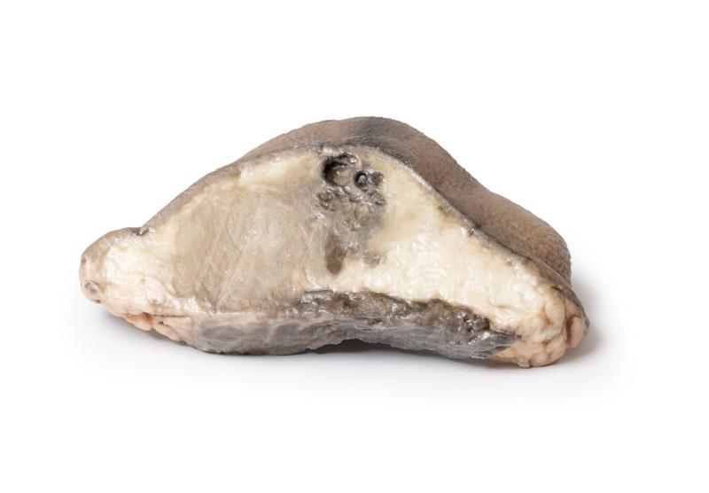
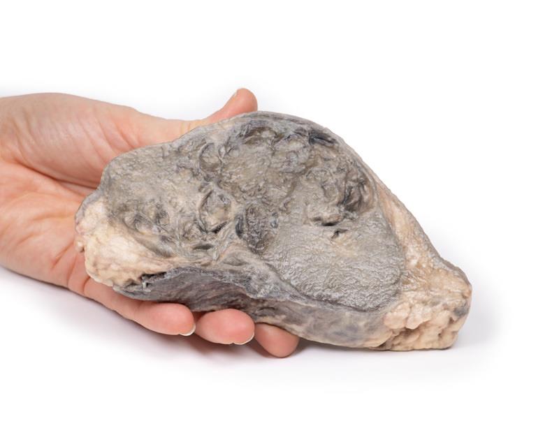
The specimen is the patient’s left breast mounted to display the cut surface. Immediately beneath and attached to the skin is a large oval tumour mass 11cm in maximum diameter. The tumour is adherent to the underlying muscle. The tumour is not encapsulated and has a variegated cut surface with areas of necrosis, haemorrhage and cyst formation. This is a breast adenocarcinoma, which involved the regional lymph nodes.
Further Information
Breast carcinoma is the second most commonly diagnosed cancer in women worldwide. It is rare in women under 30 years of age but the incidence increases significantly after 30 with the peak occurring at 70 to 80 years. Incidence has been decreasing since the introduction of breast cancer screening programmes, which offer mammography to women at risk and public awareness and education in self-examination of the breast. However, breast cancer remains one of the leading causes of cancer-related death in women.
Major risk factors for developing breast carcinoma include being of female gender (men account for 1% of breast cancer diagnosis), exposure to estrogen (early menarche, late menopause, exogenous estrogen), family history of breast cancer, being nulliparous, not breastfeeding, radiation exposure and obesity. Germline mutations in tumour suppressor genes, such as BRCA1, BRCA2, TP53, ATM, CDH1 and CHEK2 are linked with some hereditary cases of breast cancer.
Most breast neoplasms are adenocarcinomas that begin in the duct/lobular system as carcinoma in situ (DCIS). These malignancies are further subdivided according to their expression of estrogen receptors (ER) and Human epidermal growth factor 2 (HER2), which guides treatment. The most frequent sites if distant metastasis occur are bone, liver, lung and brain.
In developed countries with screening programmes most patients present following an abnormal mammogram. Symptomatic patients present with a breast mass which is classically hard, irregular, immovable. Other clinical symptoms are axillary lymphadenopathy, overlying skin changes (erythematous or thickened skin, dimpled skin (peau d‘orange) and nipple retraction. Symptoms of distant spread of disease may also cause patient presentations.
Treatment depends on the stage of the disease and the ER and HER2 status of the tumour. Surgical treatments include uni- or bilateral mastectomy or breast conserving lumpectomy. Surgical axillary node clearance is performed in cases with positive nodal disease. Radiotherapy is given to patients with high risk of local recurrence. Patients with HER2 positive cancers are treated with targeted drugs, such as trastuzumab (Herceptin). Patients with ER positive tumors can be treated using anti-estrogen therapy, such as tamoxifen. Systemic chemotherapy is also used to treat some patients with breast cancer.


