Description
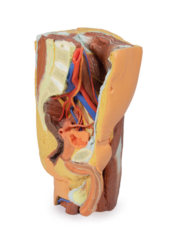
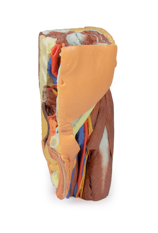
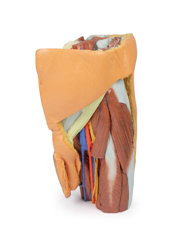
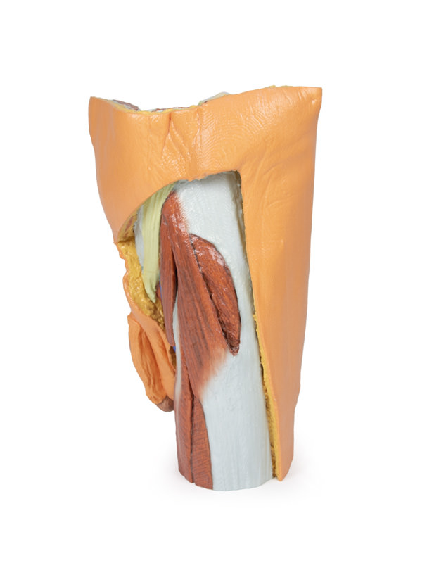
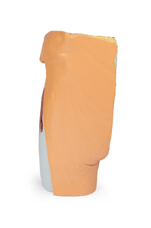
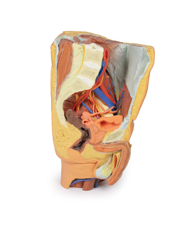
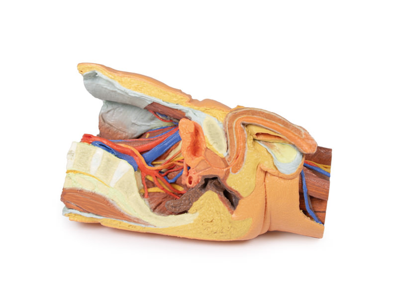
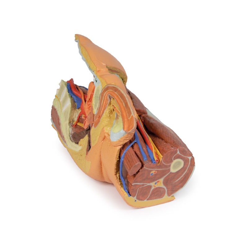
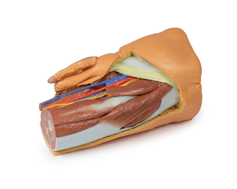
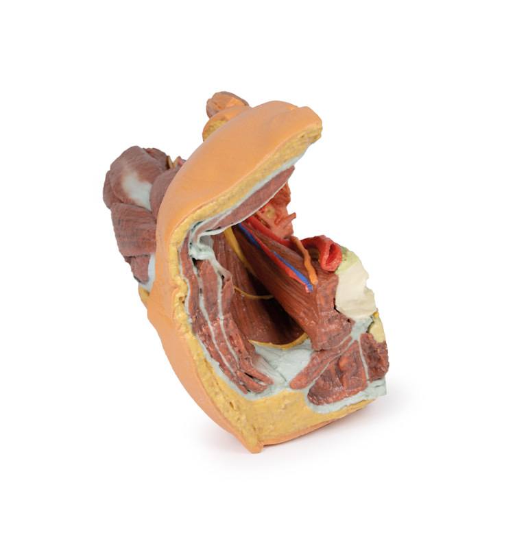
This 3D printed male left pelvis and proximal thigh (sectioned through the midsagittal plane in the midline and transversely through the L3/4 intervertebral disc) shows superficial and deep structures of the true and false pelves, inguinal and femoral region. In the transverse section, the epaxial musculature, abdominal wall musculature (rectus abdominis, external and internal abdominal obliques, transversus abdominis), psoas major and quadratus lumborum are visible and separated from each other and the superficial fat by fascial layers such as the rectus sheath and the thoracolumbar fascia. The psoas major muscle lies lateral to the external iliac artery, with the left testicular artery and vein lying on its superficial surface. More laterally (and moving inferiorly), the ilioinguinal nerve, the lateral cutaneous nerve of the thigh and the femoral nerve are positioned over the superficial surface of the iliacus muscle.
Detailed anatomical description on request.
